

Topic 56. Anatomy Part 1: Anatomy of the Lactating Human Breast
Topic 56. Anatomy Part 1: Anatomy of the Lactating Human Breast
March 19, 2024

And … we’re back! I know it is Tuesday, which is usually reserved for Pediatric Pearls, but we have test takers out there and I want to get right back into study information for them. You will be seeing two posts:
This post is the first part of the anatomy section - a review of Dr. Hartmann’s 2005 study. Anatomy Part 2 will be posted tomorrow (3/20/24) and Anatomy Part 3 on Friday (3/22/24).
I will also post Pediatric Pearls for all of you to see with no paywalls. We talk about infant formula in Pediatric Pearls - wait until you read about a jury decision in Illinois.

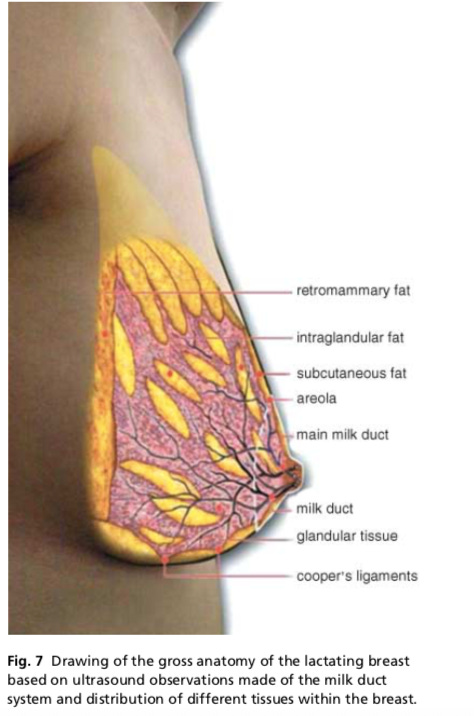
In 2005, Peter Hartmann and his team at the University of Western Australia published a study that redefined the anatomy of the lactating human breast. (Reference 1)
Link to Hartmann study This article can be downloaded at no charge.
The study used ultrasound imaging to investigate breast anatomy of 21 fully lactating women who were 1-6 months post-partum. These women were: Caucasian; between the ages of 22-38 years; with a parity of 1-4; mothers of healthy term infants; and exclusively breastfeeding.
The key findings were:
The average number of ductal openings is 9, ranging from 4-18. (This differs from the previous teaching from Gray’s Anatomy of 15-20 lobes and milk ducts in the female breast.)
All ducts branch within the areolar radius, very close to the nipple.
At milk ejection the ducts shorten and widen, increasing their diameter as the milk flows through them and returning to their resting position after the milk ejection.
Lactiferous sinuses, previously almost universally taught as present in anatomy classes and books, do not exist. From the study, “The low number and size of ducts, the rapid branching under the areola and the absence of sinuses suggest that ducts transport breastmilk, rather than store it.”
“The milk ducts at the base of the nipple were superficial, small and easily compressed. These features make them easy to occlude and difficult to palpate.”
It’s not a tidy system. The course of the ducts is convoluted, lobes are merged and sometimes overlaying.
“The distribution of adipose and glandular tissue showed wide variation between women but not between breasts within women. The proportion of glandular and fat tissue and the number and size of ducts were not related to milk production.”
The majority of glandular tissue is found within a 30-mm radius of the base of the nipple. (30 millimeters = 3 centimeters = 1.18 inches)
This is an important study to be aware of when asked questions about the anatomy of the lactating breast. Also, think of the implications of this information on the latch, hand expression, nipple shield sizing, and breast surgery.
References
Ramsay DT, Kent JC, Hartmann RA, Hartmann PE. Anatomy of the lactating human breast redefined with ultrasound imaging. J Anat. 2005;206:525-534.
Tema 56. Anatomía Parte 1: Anatomía del Pecho Humano durante la Lactancia
En 2005, Peter Hartmann y su equipo de la Universidad de Australia Occidental, publicaron un estudio que redefinió la anatomía de la mama humana en lactancia. (Referencia 1)
Hartmann Article Link - Gratis
El estudio utilizó imágenes ecográficas para investigar la anatomía de las mamas de 21 mujeres en plena lactancia que estaban entre 1 y 6 meses después del parto. Estas mujeres eran: Caucásicas; con edades comprendidas entre los 22 y los 38 años; que habían tenido de 1 a 4 partos; madres de bebés sanos a término; y con lactancia materna exclusiva.
Los resultados principales fueron los siguientes:
El número medio de aberturas ductales es de 9, con un rango de 4 a 18. (Esto difiere de la enseñanza anterior de la Anatomía de Gray de 15-20 lóbulos y conductos lácteos en la mama femenina).
Todos los conductos se ramifican dentro del radio areolar, muy cerca del pezón.
En el momento de la eyección de la leche, los conductos se acortan y ensanchan, aumentando su diámetro a medida que la leche fluye a través de ellos y volviendo a su posición de reposo después de la eyección de la leche.
Los senos lactíferos, que antes se enseñaban casi universalmente como presentes en las clases y libros de anatomía, no existen. Según el estudio, "el escaso número y tamaño de los conductos, la rápida ramificación bajo la areola y la ausencia de senos sugieren que los conductos transportan la leche materna, en lugar de almacenarla".
"Los conductos lácteos de la base del pezón eran superficiales, pequeños y se comprimían fácilmente. Estas características los hacen fáciles de ocluir y difíciles de palpar".
No es un sistema ordenado. El curso de los conductos es enrevesado, los lóbulos están fusionados y a veces se superponen.
"La distribución del tejido adiposo y glandular mostró una amplia variación entre las mujeres, pero no entre las mamas de una misma mujer. La proporción de tejido glandular y adiposo y el número y tamaño de los conductos no estaban relacionados con la producción de leche."
La mayor parte del tejido glandular se encuentra en un radio de 30 milímetros de la base del pezón. (30 milímetros = 3 centímetros = 1,18 pulgadas)
Este es un estudio importante que hay que tener en cuenta cuando se hacen preguntas sobre la anatomía de la mama en lactancia. También hay que pensar en las implicaciones de esta información en el agarre, la extracción manual, el tamaño del protector del pezón y la cirugía mamaria.
Referencias
Ramsay DT, Kent JC, Hartmann RA, Hartmann PE. Anatomy of the lactating human breast redefined with ultrasound imaging [Anatomía de la mama humana en lactancia redefinida con imágenes ecográficas.]. J Anat. 2005;206:525-534.